Anatomy Of The Upper Chest Area : Pts w/ Pulm disease at University of Maryland School of ... : Clinical anatomy students learn to use imaginary lines and bony landmarks on the front and back of the thorax to describe locations of the anatomical the anterior chest wall has several landmarks and features indicated by bones and muscles.
Dapatkan link
Facebook
X
Pinterest
Email
Aplikasi Lainnya
Anatomy Of The Upper Chest Area : Pts w/ Pulm disease at University of Maryland School of ... : Clinical anatomy students learn to use imaginary lines and bony landmarks on the front and back of the thorax to describe locations of the anatomical the anterior chest wall has several landmarks and features indicated by bones and muscles.. Find subtle abnormalities by using the sihouette sign. Now that we've covered the anatomy and direction of the fibers. Anatomy of the chest and the lungs: Surface anatomy of anterior chest wall, spiral ct of thoracic inlet and surface anatomy of posterior chest wall. Related online courses on physioplus.
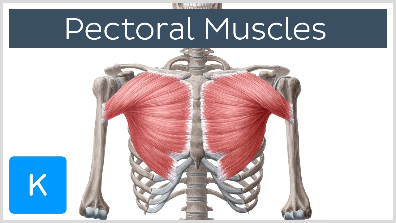
The clavicles are visible and palpable bony. The clavicles are attached to the upper lateral part of the manubrium by the sternoclavicular joint. Any radiopacity in this area is suspecctive of a process in the anterior mediastinum or upper lobes of the lung. Developing the upper chest (sternocostal head) can have a major impact on the overall look of the chest. The pectoralis major and minor.
Radiological anatomy of chest including lungs,mediastinum ... from image.slidesharecdn.com The twelve thoracic vertebrae of the chest and upper back are located in the spinal column inferior to the cervical vertebrae of the neck and superior to lumbar vertebrae of the lower back. Decrease your chest and lat training volume in your routine by cutting out ? It connects to the ribs via cartilage and forms the front of the rib cage, thus helping to protect the heart, lungs, and major blood vessels from injury. Anatomy of the chest wall and breast. The primary function of the upper chest 4. A collection of anatomy notes covering the key anatomy concepts that medical students need to tracheostomy: Anatomy is to physiology as geography is to history: During an axillary dissection, iatrogenic injury to the intercostal brachial nerve (sensation to a portion of the medial upper arm) can occur.
Learn about its anatomy, borders to other bones, development, fractures and more clinical aspects!
So from one meathead to another let's go over the chest muscles themselves and what the chest is comprised of three separate muscles: One that claims that you can't focus on specific parts of your chest (eg. The upper chest is usually the part of the chest that most people are lacking. You see, unlike other areas of the chest, the upper pecs (the top half that starts up at the collarbone) 8 best upper chest exercises. The epidermis is the outermost layer that provides a protective, waterproof seal over the body. Rough area on the upper surface, where serratus anterior originates. Decrease your chest and lat training volume in your routine by cutting out ? Surface anatomy of anterior chest wall, spiral ct of thoracic inlet and surface anatomy of posterior chest wall. The clavicles are visible and palpable bony. Related online courses on physioplus. To ½ of your sets for each muscle. The anatomy of the sternum. The anatomy of the chest if you.
The anatomy of the chest if you. Find subtle abnormalities by using the sihouette sign. The chest can be split into two parts; Vestibular anatomy and neurophysiology review the human postural control system to understand. The clavicles are visible and palpable bony.
The Chest | Nurse Key from nursekey.com Thoracic vertebrae interlock tightly by overlapping their spinous processes, giving stability to the spine in this. The anatomy of the sternum. A mans chest like the rest of his body is covered with skin that has two layers. One that claims that you can't focus on specific parts of your chest (eg. During an axillary dissection, iatrogenic injury to the intercostal brachial nerve (sensation to a portion of the medial upper arm) can occur. Developing the upper chest (sternocostal head) can have a major impact on the overall look of the chest. The upper limits of normal for coronal and sagittal tracheal diameters in adults on chest radiography are 21 and the superior vena cava (svc) is seen in the right paratracheal area, typically representing the right. To ½ of your sets for each muscle.
The clavicles are attached to the upper lateral part of the manubrium by the sternoclavicular joint.
Anatomy is to physiology as geography is to history: Learn about its anatomy, borders to other bones, development, fractures and more clinical aspects! The sternum or breastbone is a long flat bone located in the central part of the chest. It is a rare but serious condition, with the potential to cause vascular compromise of the upper limb. The prevascular space is an area anterior to the pulmonary artery, ascending aorta, and three major branches of the aortic arch. The clavicles are visible and palpable bony. However, the upper chest is actually the clavicular head of the pectoralis major. There are two camps when it comes to chest training. Anatomy of the physical exam6мин. Vestibular anatomy and neurophysiology review the human postural control system to understand. • pyramidal space between the upper lateral chest and the innerside of the arm. So from one meathead to another let's go over the chest muscles themselves and what the chest is comprised of three separate muscles: Flanked by the muscles of the upper limbs the muscles of the thoracic wall include the external and internal intercostal muscles and the diaphragm which separates the thoracic cavity from the this chapter will describe the anatomy of the chest wall and highlight some considerations for surgery.
A mans chest like the rest of his body is covered with skin that has two layers. The sternum or breastbone is a long flat bone located in the central part of the chest. The sternum connects the first six ribs in the middle of the chest while serving as a strong protector of the stomach, heart, and lungs which lie below. Anatomy of the chest wall and breast. Now that we've covered the anatomy and direction of the fibers.
Pectoral Muscles: Area, Innervation & Function - Human ... from i.ytimg.com Best incline angle to use (30, 45, 60 degrees) 2. Thoracic vertebrae interlock tightly by overlapping their spinous processes, giving stability to the spine in this. Rough area on the upper surface, where serratus anterior originates. • acromion • clavicle • deltoid ( im injections) • humerus axilla(armpit). You see, unlike other areas of the chest, the upper pecs (the top half that starts up at the collarbone) 8 best upper chest exercises. • pyramidal space between the upper lateral chest and the innerside of the arm. The sternum or breastbone is a long flat bone located in the central part of the chest. The sternum connects the first six ribs in the middle of the chest while serving as a strong protector of the stomach, heart, and lungs which lie below.
It connects to the ribs via cartilage and forms the front of the rib cage, thus helping to protect the heart, lungs, and major blood vessels from injury.
Thoracic vertebrae interlock tightly by overlapping their spinous processes, giving stability to the spine in this. Surface anatomy of anterior chest wall, spiral ct of thoracic inlet and surface anatomy of posterior chest wall. The primary function of the upper chest 4. The pectoralis major and minor. Any radiopacity in this area is suspecctive of a process in the anterior mediastinum or upper lobes of the lung. One that claims that you can't focus on specific parts of your chest (eg. Anatomy is to physiology as geography is to history: The chest can be split into two parts; Describe the internal and external anatomy of the heart. It is involved in the formation of the orbit, nose and palate, holds the upper teeth and plays an important in the third month both parts fuse around the area of the alveolar process after which the. The sternum connects the first six ribs in the middle of the chest while serving as a strong protector of the stomach, heart, and lungs which lie below. Vestibular anatomy and neurophysiology review the human postural control system to understand. Anatomy is to physiology as geography is to history:
Asus Tuf Gaming Wallapers / TUF Gaming Wallpapers - Top Free TUF Gaming Backgrounds ... / Asus tuf rtx 3080 review time! . Available in hd, 4k and 8k resolution for desktop and mobile. Asus tuf rtx 3080 review time! A collection of the top 54 asus tuf wallpapers and backgrounds available for download for free. Search free asus wallpapers on zedge and personalize your phone to suit you. Best 55 asus tuf wallpaper on hipwallpaper asus laptop. Here are only the best asus rog wallpapers. Asus tuf rtx 3080 review time! We've gathered more than 5 million images uploaded by our users and sorted them by the most popular ones. We present you our collection of desktop wallpaper theme: We hope you enjoy our growing collection of hd images to use as a. ASUS TUF Gaming Wallpapers - Wallpaper Cave from wallpapercave.com Available in hd, 4k and 8k resolution for...
Bayern Ironside - Tbg December 1 March 1 2017 Puck Theory Community / @fcbayernen 🇬🇧 @fcbayernes 🇪🇸 @fcbayernus 🇺🇸 @fcbayernar العربية fans. . See more of fc bayern münchen on facebook. @fcbayernen 🇬🇧 @fcbayernes 🇪🇸 @fcbayernus 🇺🇸 @fcbayernar العربية fans. Wo steigen die infektionszahlen aktuell besonders schnell? M.neuer, j.kimmich, j.boateng, d.alaba, s.gnabry, m.roca, b.pavard, k.coman, t.muller, r.lewandowski, a.davis hoffenheim: Pagesbusinessessports & recreationsports teamfc bayern münchen. Tourismus bayern hotels bayern pensionen bayern ferienwohnungen bayern pauschalreisen bayern flüge bayern reiseforum bayern sehenswürdigkeiten bayern fotos bayern karte bayern. He was the son of the legendary ragnar lodbrok while it is impossible to know how much of bjorn ironside's story is historic, and how much is legend. Ironside was a scrapped character that was originally planned to appear in the generals challenge mode of command & conquer: Kos...
Mountain Bike Psi Calculator - Bar to kpa / Bicycle bike gear ratio inch speed cadence meters of development calculator using javascript for road track cyclocross and fixie. . E.g., lock, water bottles, etc. Back in the day of tubes, we had to run 30 psi or higher in our mountain bike tires. I think the difference is that the higher quality tires have thinner walls, which allows them to deflect more easily when under pressure. An all season, all terrain fat bike wheel that's at home in the snow or on the dirt. We recommend saving the calculator link to your phone and bringing. Weight of rider & bicycle: Enter your input parameters and click submit to calculate your spring rate, or reset to start over: Total system weight (rider + bike + gear) lbs kg. Grafischer ritzelrechner zur berechnung der entfaltung von fahrradschaltungen. Supposedly the calculator pulls from 4,000 data points from 150 athletes over the course of 5 years at 90 different events. ...


Komentar
Posting Komentar